For decades, noise-induced and age-related hearing loss research efforts have focused on the loss of hair cells and the threshold elevations this causes. Hair cells have long been considered the most vulnerable elements in the inner ear, but researchers working at the Massachusetts Eye and Ear Infirmary’s (MEEI) Eaton Peabody Laboratory at Harvard Medical School have now shown that nerve fibers are even more vulnerable to damage.
At the 167th meeting of the Acoustical Society of America (ASA), held last week (May 5-9) in Providence, RI, the researchers reported their discovery of “hidden hearing loss.”
How Hidden Hearing Loss Occurs
In the normal ear, sound waves are transmitted through the middle ear bones to the inner ear, where they cause vibrations in the sensory epithelium called the “organ of Corti.” The organ of Corti turns this mechanical function into electrical pulse trains in the fibers of the cochlear nerve, which then carries the information to the brain for analysis of the acoustic scene.
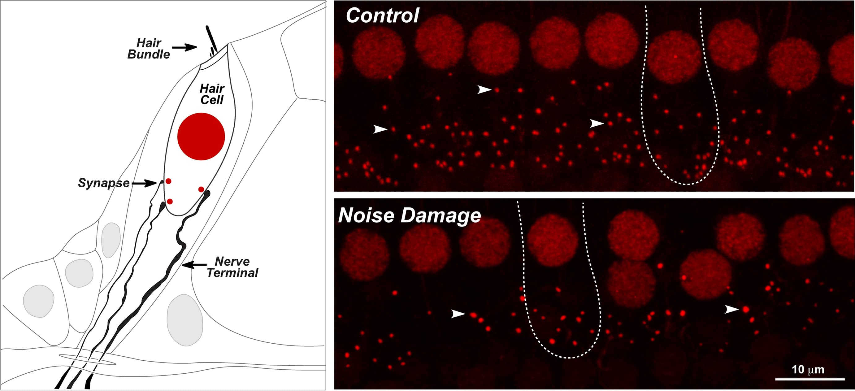
(Click on graphic to enlarge.) The left figure shows an inner hair cell, with its hair bundle at one end and with 3 of its roughly 20 synapses with cochlear nerve terminals at the other end. The cell’s nucleus is shown in red, and each synapse is marked by a smaller red dot. Immunostain recognizes a molecule present in the nucleus and also in a key synaptic component within the hair cell, called a “ribbon.” The images shown on the right were taken with a confocal microscope. The upper image shows 8 inner hair cells from a normal ear (each cell has one large red nucleus) and about 150 small red dots (arrowheads), each of which shows a synapse between a hair cell and a cochlear nerve fiber. The bottom image shows another ear a few days after exposure to a noise that caused only a transient threshold elevation. The confocal image shows no loss of hair cells, but a striking loss of synapses, which will never recover.
“The organ of Corti contains two types of sensory cells: outer and inner hair cells,” explained Charles Liberman, director of MEEI’s Eaton Peabody Laboratory. “The sensory cells are called ‘hair cells’ because of the hair-like tufts of microvilla on their apical surfaces, which are called ‘stereocilia.’ Bending the stereocilia opens ion channels in the nerve hair cells and allows a current to flow that ultimately excites the fibers of the cochlear nerve. This is the heart of the mechanical-to-electrical transduction process in the inner ear.”
How did hidden hearing loss remain “hidden” so long? There are two key reasons, according to Liberman.
“First, the field of auditory neuroscience didn’t appreciate until recently that you can lose up to 90% of your cochlear nerve fibers without a change in the ability to detect a tone in quiet,” he said. “Tone detection in quiet is the basis of the threshold audiogram—the gold standard test of hearing function. The fact that thresholds may transiently elevate and then recover within hours or days after an acoustic overexposure doesn’t mean that the inner ear has recovered.”
Second, the most vulnerable part of the neuron turns out to be the synapse between the nerve terminal and the hair cell—and it happens to be difficult to see. “Until recently,” said Liberman, “they could only be seen and counted by using an electron microscope.”
To maneuver around this, the researchers stained synapses with antibodies that target molecular structures within the synapse, which allowed the synapses to be seen and easily counted in a light microscope. This enabled them to view a large number of synapses on hair cells in a normal ear, as well as the greatly reduced numbers of synapses hair cells following a noise exposure that caused only a transient elevation of thresholds. Liberman and colleagues counted synapses on thousands of hair cells.
“Each missing synapse represents a cochlear nerve fiber that has been disconnected due to retraction of the terminal segment—it will never reconnect,” Liberman noted. “It no longer responds to sound, and, within a few months or years, the rest of the neuron will disappear.”
Sober Implications for Public Health
The public health implications of this finding are quite sobering. “All of our federal noise exposure guidelines are based on the assumption that noise-exposures causing only transient threshold elevation are benign,” Liberman points out. “That assumption is almost certainly unwarranted.”
Liberman and colleague Sharon Kujawa, director of audiology for MEEI, demonstrated noise-induced hidden hearing loss in three mammalian ears: mouse, guinea pig’ and chinchilla. “There is every reason to believe the same phenomenon applies to human ears,” he said.
This suggests that every time we go to loud concerts or use power tools without ear protection, we may be losing cochlear nerve fibers and increasing our degree of hearing impairment.
Holding out hope that the damage may possibly be reversible, Liberman is working with Gabriel Corfas, senior associate in neurology and otolaryngology for Boston’s Children’s Hospital, to explore potential therapies.
“Since the ultimate death of neural cell bodies and axonal projections to the brain is so slow, there is an extended therapeutic window where delivery of chemicals to elicit sprouting of the nerve terminals might be able to reestablish synaptic connections between neuron and hair cell,” Liberman noted.
A class of secreted proteins called neurotrophins has shown to play a key role in the survival of cochlear nerve fibers, and Liberman and Corfas recently found that overexpression of neurotrophin, in a genetically modified mouse line, partially rescued the post-exposure synaptic loss. “Our work is ongoing in this area,” he said.
Last week, The Hearing Review also reported on research in which cochlear implants stimulate neurotrophin production and thereby regenerate nerve fibers.
Source: Acoustical Society of America via Newswise.com