Otoscopy using a binocular microscope (referred to as otomicroscopy) represents a serious upgrade for hearing care professionals who are commited to providing superior cerumen management and hearing aid services. Moreover, when clinicians include video otomicroscopy, they add a powerful tool for patient education and motivation, as well as data storage for patient records.

|

|
| Gene R. McHugh, EdD, is an audiologist and owner of Colorado Springs Audiology Inc, Colorado Springs, Colo; and Robert M. Traynor, EdD, is an audiologist and owner of Audiology Associates Inc, Greeley and Johnstown, Colo. | |
In a typical audiology practice, it is estimated one in four patients presents with varying degrees of cerumen impaction that should be addressed prior to examination and/or hearing aid services.1 With age, prevalence of cerumen problems tends to increase,2 with reports of cerumen impaction in nursing home residents as high as 65%.3,4
According to a recent consortium on cerumen management,5 cerumen is considered “problematic” when it causes otologic or audio-vestibular symptoms, prevents necessary assessment of the ear including audiologic assessment, or causes problems for persons wearing hearing aids. As a method of cerumen removal, binocular microscopy is considered superior primarily due to the improvement in overall sight, especially with regard to depth perception.6 For many years, our otolaryngology colleagues have been well aware of the value of operating microscopes; possessing at least one in-office microscope in an ENT practice is practically axiomatic.7,8
Interestingly, trends toward using binocular microscopes are also being realized in other professions. For example, in the last 10 years, there has been a wide range of articles in dental journals making convincing arguments regarding the value of operating microscopes in dental offices. The trend of using dental microscopes initially started in the specialties of endodontics and periodontics. Recent articles suggest the number of general dentists using dental operating microscopes (DOMs) is steadily growing for restorative procedures.9-11
Cerumen management is within the scope of practice for most audiologists in the United States, although a few state licenses specifically exclude cerumen management by audiologists.12-14 For purposes of removing cerumen, most clinicians were trained with, and continue to use, otoscopic instruments other than microscopes.15,16
In our opinion, otoscopy using a binocular microscope (referred to as otomicroscopy) represents a significant upgrade for hearing care professionals who are commited to providing superior cerumen management and hearing aid services.7 Moreover, when clinicians include video otomicroscopy, they add a powerful tool for patient education and motivation, as well as data storage for patient records.

|

|
| FIGURE 1. Binocular microscopy improves overall sight, especially illumination and depth perception. | FIGURE 2. Gene McHugh, who has been using otomicroscopy for over 15 years, examines a patient. |
Advantages of Binocular Microscopy
Binocular microscopy is superior as a cerumen management tool because of the significant improvement in sight. Microscopes can magnify vision up to 20X and utilize an intense “co-axial” fiber-optics light to illuminate the ear canal (Figure 1). Most importantly, binocular microscopes provide 3-dimensional vision and, as a result, offer exceptional depth perception. In comparison, traditional otoscopy and video otoscopy provide only a 2-dimensional view.
The video otomicroscope is mounted on a stable and adjustable arm, which allows for nearly hands-free manipulation during otoscopic examinations and cerumen removal procedures (Figure 2). Due to the improvement in vision and ability to use both hands during procedures, cerumen removal using otomicroscopy becomes easier for the clinician when manually removing cerumen and thus safer for the patient. For this reason, otomicroscopy is considered “state of art” as a tool for cerumen management.17,18
|
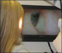
| FIGURE 3. The patient views the process of cerumen removal. Most are fascinated by this procedure. |
|
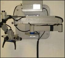
| FIGURE 4. Wall-mount binocular microscope with wire covers. |
Perceived Obstacles
According to McHugh19 and others,18,20 the “perceived” reasons for not using a microscope in an audiology practice include: 1) the relatively high initial purchase price; 2) little or no return on investement due to lack of direct income from cerumen management services; 3) insufficient clinic space in one’s office, and 4) little or no training to become proficient using otomicroscopy. We will address these issues individually.
Cost. Initial costs are high, but not out of the range of most private practice audiologists. Part of the perception that microscopes are out of the financial reach for audiologists is created by costly operating room microscopes, which can be in the range of $25,000 to $250,000. However, otomicroscopes designed for audiologists are in the range of $4,000 to $7,000. The inclusion of a color video camera (sometimes referred to as a charge-coupled device or CCD) will add another $3,000-$4,000 to the cost.
Many audiologists have found video-otoscopy beneficial in terms of patient relations and education. Video otomicroscopy replaces this equipment with an instrument that is more useful and offers better capability to patient care. The final touch in implementing video otomicroscopy in one’s practice is purchasing a TV or LCD monitor with good resolution. These monitors can be as basic as a simple 17-inch color TV (costing approximately $150) or as complex as a 32-inch HDTV (approximately $1,200). Regardless of the size/quality of the monitor, those involved with the clinic will recognize the value of this investment.
Return on investment. Personal experience has shown there to be a substantial “return on investment” by implementing video otomicroscopy, especially for the purposes of cerumen management. Unlike many “must have” pieces of equipment that eventually sit in a corner of the office, the video otomicroscope will be an essential part of the audiological armamentarium for many years as shown by trends in ENT and other professions.19 Regardless of minimal reimbursement specifically related to cerumen management services, the authors believe that the intangible improvement in professionalism and quality of care offered by video otomicroscopy is well worth the investment. Finally, including a video camera with constant visual feedback is “key” to attaching a tangible value for patients, family members, and office auxiliaries so they can see what the clinician sees (Figure 3).21
Space limitations. While most video otomicroscopes are mounted on a floor stand and take up space in the treatment room, microscope units can be conveniently wall-mounted to be up and out of the way (Figure 4). This allows for other devices to be stored in the room and the systems provide more room to work. While a floor-mount scope can be moved from room to room, the base is relatively large, cumbersome, and difficult to move, so it tends to stay in the same room taking up valuable office space. For this reason, wall-mount units offer a design alternative to reduce clutter in the treatment room.
It is necessary to account for all of the electrical wiring for the scope and video accessories. Reputable microscope companies offer trained technicians who will secure the scope to the wall.
Training/learning issues. Like any new piece of equipment, the system is of limited or no benefit without proper introduction, orientation, and customization by the manufacturer’s representative. For audiologists with academic and practical knowledge of the ear canal and its anatomy, clinical technique in viewing the ear canal and tympanic membrane develops rather easily. Although it is possible to self-train for the use of otomicroscopy or video otomicroscopy, taking a short introductory class can speed up the learning curve. Within a few weeks, the microscope should become comfortable to use, and within a few months the skill level in safely removing cerumen using manual or suction maneuvers is vastly improved.
Microscope Distributors
Currently, there are four microscope distributors marketing to audiologists: Jedmed, Prescott’s, SBCS Medical, and Seiler. Other companies offering ENT-style microscopes appropriate for audiological purposes include Zeiss, Global Surgical Corp, Leica Microsystems, and Alltion (China), along with suppliers who will sell their microscopes online. Most offer basic floor and wall-mount microscopes with a wide range of prices and service plans after the product is delivered. Obtain as much information as possible before selecting where to purchase your microscope. There are differences in the quality of microscopes, with some being easier to adapt to than others. Other purchasing considerations include the provision of on-site installation, equipment customization, and basic sales and service staff. As with all audiometric equipment, long-term service and support should be an important component in your final decision.

|

|
| FIGURE 5. The Medicap system used in video data storage. | FIGURE 6. The various working components of the video otomicroscope. |
Ancillary Equipment
Examination chair. To obtain the maximum benefit from video otomicroscopy, it is necessary to obtain a high-level examination chair. The chair needs to be height adjustable with swivel ability for correct patient positioning. A headrest is also essential for stabilizing the patient’s head during video otomicroscopy procedures. A basic fixed upright examination chair that manually swivels and can be adjusted for height with a foot pump lever costs approximately $1,000-$1,500. More sophisticated chairs with electronic adjustments and a recline option are available for about $3,000-$6,000.
Data storage. A data storage device that will capture still pictures and include video recordings of various procedures is a popular addition. It is best to inquire with a representative from a microscope company to determine your needs and the special equipment required for data storage.
One option for data storage is the Medicap-USB200 (offered by Medi-capture and available from Prescott’s and Jedmed) at a cost of around $4,000. The costs and features in various video recording, as with most computer and video-related equipment, can range widely.

|

|
| FIGURE 7. Adjusting one of locking wheels on the microscope arm to prevent drift. | FIGURE 8. 12.5X magnification lenses. Each lens has ± diopter adjustment. |
Getting Familiar with the Basic Parts of an Operating Microscope
|
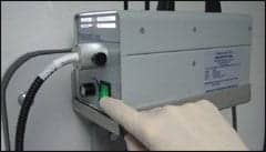
| FIGURE 9. Fiber optics light source with variable gain. |
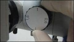
|
| FIGURE 10. Magnification control. Ranges for 7.5X to 20X. |
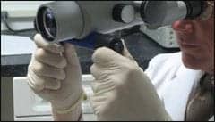
|
| FIGURE 11. The interchangeable objective lens defines the working distance between microscope and patient’s ear. |
Figure 6 shows the major working components of a video otomicroscope, including:
- Adjustable arm and coupling
- Eyepieces and binocular lenses
- Fiber-optics light source, cable, and fiber-optic block
- Magnification changer
- Focusable objective lens
- Handles
Adjustable arm and coupling. Correct installation of wall-mounted microscopes is important to avoid uncontrollable drifting.22 Assuming the unit has been installed correctly, the microscope’s arm should be easy to position, and once in place, hold the microscope stable and without drift. The body of the microscope can be positioned from 30° to 90°, depending on if the patient is lying down or in a sitting position. There are knobs (locking wheels) at strategic points along the arm to hold the scope in the correct position during the examination and/or procedures.
Eyepieces and binocular lenses. Each eyepiece has a lens with 12.5X magnification. Lenses can be widened or narrowed depending on inter-pupillary distance. Adjust the diopter on the lenses for each eye if there are vision differences between eyes. If vision is relatively equal, set both lenses to the normal level (ie, 0 diopters). If you work in an office with multiple users, make a note of the diopter settings for each eye of each user for future reference.
Fiber-optics light source, cable, and fiber-optic block. In operating microscopes, a variable fiber-optics light is projected through a cable that focuses the beam of light directly into the ear canal. As a result, when the microscope is repositioned, the light remains on the focused object. Clinicians who use optical loupes for cerumen management are aware that, as magnification increases, more light is required into the ear canal to compensate for the magnified view. This is the reason loupes with higher magnification require stronger and more direct lighting to adequately illuminate the ear canal.
Magnification changer. As indicated previously, each binocular eyepiece provides 12.5X magnification. A manual control on the side of the microscope’s body will alter that magnification by a factor 0.6X, 1.0X, or 1.6X. As a result, there are magnification levels of 7.5X, 12.5X, and 20X. For most cerumen management applications, otomicroscopy is best performed at 7.5X magnification, occasionally at 12.5X magnification, and rarely at 20X magnification. As a reference, the normal tympanic membrane is 10 mm (about 1/3 inch). At 20X magnification, the tympanic membrane looks like it is 200 mm (8 inches).
Focusable objective lens. The objective lens (on the end of the microscope tube) defines the working distance between microscope and the desired focus area. For example, on the Prescott’s OMNI-10 binocular microscope shown in these photos, objective lenses come in four sizes: 175 mm (approximately 7 inch), 200 mm (or 8 inch), 225 mm (or 9 inch) and 250 mm (or 10 inch). Clinicians with shorter arm lengths should use the 175 mm or 200 mm objective lenses. Clinicians with longer arms will likely prefer the 225 mm or 250 mm objective lens for more working distance. It is well advised to check with the microscope’s representative to determine the objective lens that is ergonomically best for the clinician.
Handles. Handles are located on either side of the microscope’s body to freely move the microscope into various position as necessary to see different parts of the canal.
In conclusion, clinicians experienced in cerumen management should find it relatively easy to transition to otomicroscopy and may even find it rewarding and quite enjoyable.
Acknowledgement
Gene R. McHugh is a consultant with Prescott’s.
References
- Gleitman RM, Ballachanda BB, Goldstein DP. Prevalence of cerumen impaction in the general population. Hear Jour. 1992;45(5):28-32.
- Roeser RJ, Ballachanda BB. Physiology, pathophysiology, and anthropology/epidemiology of human ear canal secretions. J Am Acad Audiol. 1997;8:391-400.
- Freeman RB. Impacted cerumen: How to safely remove earwax in an office visit. Geriatrics. 1995;50:52-53.
- Lewis-Cullinan C, Janken JK. Effect of cerumen removal on the hearing ability of geriatric patients. J Adv Nurs. 1990;15:594-600.
- Roland PS, Smith TL, Schwartz SR, Rosenfeld RM, Ballachanda B, Earll JM, Fayad J, Harlor AD, Hirsch BE, Jones, SS, Krous HJ, Magit A, Nelson C, Stutz DR, Wetmore S. Clinical practice guideline: Cerumen impaction. Otolaryngol– Head Neck Surg. 2008;139:S1-S21.
- McCarter DF, Courtney AU, Pollart SM. Cerumen impaction. Am Fam Physician. 2007;75:1523-1528.
- Dohlman GF. Olaf Nylen and the birth of the otomicroscope and microsurgery. Arch Otol. 1969;90:161-165.
- Saunders WH. The operating microscope as an office instrument. Arch Otolaryngol. 1980;106(7):444.
- Nase JB. Clinical operating microscopes; They’re not just for endodontists anymore. Pa Dent J. 2005;Sept-Oct:30-33.
- Maloof D. Practical implementation of a microscope into general practice. Dental Practice, March/April 2006:124-126.
- Mamoun JS. A rationale for the use of high-powered magnification or microscope in general dentistry. Gen Dentistry. 2009;Jan/Feb:18-26.
- American Speech-Language-Hearing Association. External auditory canal examination and cerumen management. Asha. 1992;34:22-24.
- American Academy of Audiology. Scope of practice. Available at: www.audiology.org/resources/documentlibrary/Pages/ScopeofPractice.aspx. Accessed January 31, 2009.
- Schneider N. Motion to limit CM by audiologists passed at a public session dated January 12, 2006. New Jersey Audiology and Speech-Language Advisory Committee. Available at: www.nj.gov/oag/ca/aud/minutes/aud112.htm. Accessed January 29, 2009.
- Roeser R, Roland P. What audiologists must know about cerumen and cerumen management. Am J Audiol. 1992;1:27-35.
- Chaiken R. Cerumen Management. Available at: www.audiologyonline.com/articles/article_detail.asp?article_id=215. Accessed January 31, 2009.
- Ballachanda B. The Human Ear Canal: Theoretical Considerations and Clinical Applications Including Cerumen Management. San Diego: Singular Publishing Group; 1995.
- Purdy J. Managing cerumen. Available at: www.audiologyonline.com/articles/article_detail. asp?article_id=365. Accessed May 15, 2008.
- McHugh E. Cerumen management with otomicroscopy. Article 2064. Available at: www.audiologyonline.com/articles/article_detail.asp?article_id=2064. Accessed January 31, 2009.
- Clark D. The big push to clinical microscopes for esthetic dentistry. Contemporary Esthetics Restorative Practice. 2005;Nov:30-34.
- Clark D. Maximizing the return on investment of an operating microscope. Dental Economics. 2004;May:66-75.
- Clark D. A glance at what to look for when purchasing a new microscope. Dentistry Today. 2005; August [Buyer’s Guide]:100-104.
Correspondence can be addressed to HR at [email protected] or Gene McHugh at .
Citation for this article:
McHugh G, Traynor R. Video Otomicroscopy: A New “Vision” in Cerumen Managem Hearing Review. 2009;16(5):14-22.


