The system aims to provide insight into sensory hair cell development and potential pathways for regeneration.
Researchers at the University of Miami Miller School of Medicine are developing an organoid-on-a-chip system to model the development of the human inner ear and explore how damaged sensory hair cells might be regenerated. The project recently received an approximately $2.5 million, five-year RO1 grant from the US National Institutes of Health (NIH).
The research seeks to recreate the spatial division that occurs during embryogenesis, where molecular signals guide the formation of the cochlea and vestibular system.
“Our project will provide invaluable insight into how the hair cells responsible for hearing and balance develop and, potentially, how to regenerate them,” says Pei-Ciao Tang, PhD, a research assistant professor in the Miller School’s department of otolaryngology — head and neck surgery. “A glimpse of this process could contribute to new therapeutic strategies for hearing loss and vestibular disorders.”
Mimicking Embryonic Chemical Gradients
The system, designed in collaboration with the lab of Ashutosh Agarwal, PhD, an associate professor of engineering at the University of Miami, uses a microscale device to hold a human stem cell-derived organoid. The organoid is positioned between two parallel microfluidic channels, each delivering a distinct chemical environment to replicate the morphogen gradients that partition the inner ear during fetal development.
“On the basic level, the challenge was how do you fool an organoid into believing it’s part of a growing organism?” says Dr Agarwal. “The answer is by exposing it to two very different chemical environments, which is exactly what happens in development.”
One channel supplies morphogens intended to promote cochlear development, while the other allows the organoid to default toward a vestibular fate. A key goal for the team is to reliably induce the formation of cochlear tissue, which has historically been more difficult to generate from stem cells than vestibular tissue.
Pursuing the Possibility of Regeneration
While studying cochlear development, the team will use the more accessible vestibular system model to investigate sensory hair cell regeneration. Unlike the cochlea, where hair cell loss is permanent, some evidence suggests the vestibular system has a limited capacity to regenerate these cells.
The researchers will explore the epigenetic mechanisms, such as DNA methylation, that may limit this regenerative process. Dr Tang will map chromatin accessibility and gene expression in the organoids, including after exposure to an ototoxic aminoglycoside antibiotic, to identify developmental pathways involved in injury response. Collaborator Derek Dykxhoorn, PhD, professor in the Dr John T Macdonald Foundation department of human genetics, will then use CRISPR-based tools to test candidate genes.
“If we can identify the key pathways that drive hair cell fate, we may be able to enhance hair cell regeneration by modulating them,” says Dr Dykxhoorn. “This would give us targets for therapeutic approaches to address disorders of the inner ear.”
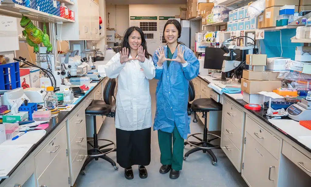
Featured image: Dr. Pei-Ciao Tang (left, with Dr. Diane Jung) is studying how hair cells develop, and how to regenerate them. Photo: University of Miami Miller School of Medicine