A new one-step fabrication method improves the design of flexible 3D microelectrode arrays for neural applications.
Neural interfaces are crucial in restoring and enhancing impaired neural functions, but current technologies struggle to achieve close contact with soft and curved neural tissues. Researchers at Pusan National University have introduced an innovative method—microelectrothermoforming (μETF)—to create flexible neural interfaces with microscopic three-dimensional (3D) structures. Their findings show how this method improves neural recording and stimulation, with potential applications in cochlear implants and brain-computer interfaces.
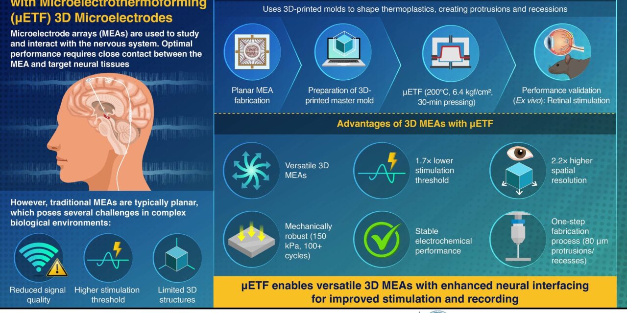
Microelectrode arrays (MEAs) are widely used for recording brain activity and stimulating neural tissues. However, conventional MEAs are typically flat, limiting their ability to conform to the natural curves of neural structures. Existing methods for adding 3D features require multiple fabrication steps, increasing complexity and restricting design possibilities.
Study Inspiration
To overcome these limitations, a team led by Associate Professor Joonsoo Jeong and Associate Professor Kyungsik Eom developed μETF, inspired by plastic thermoforming, a common technique for molding plastic sheets into different shapes. Their findings are published online on January 22, 2025, in the journal of npj Flexible Electronics. “The idea for this study came from a simple observation of plastic lids on take-out coffee cups. I realized that this plastic forming method could be applied at a microscopic level to create 3D structures for neural electrodes,” says Dr Jeong.
Method
The μETF method involves heating a thin, flexible polymer sheet embedded with microelectrodes and pressing it against a 3D-printed mold. The researchers used liquid crystal polymer (LCP) as the substrate due to its mechanical strength, biocompatibility, and long-term stability. This process forms precise protruding and recessed structures, enhancing the electrode’s proximity to target neurons while preserving its electrical properties. Unlike traditional micromachining approaches, μETF simplifies fabrication and allows for a wide range of complex 3D structures, including wells, domes, walls, and triangular features, all within a single MEA.
μETF Applications
In a proof-of-concept study, the researchers applied μETF to develop a 3D MEA optimized for retinal stimulation in blind patients. Computational simulations and lab experiments showed that the 3D electrodes reduced stimulation thresholds by 1.7 times and improved spatial resolution by 2.2 times compared to traditional flat electrodes. “Our 3D structures bring the electrodes closer to target neurons, making stimulation more efficient and precise,” Dr Eom explains.
The researchers see μETF being used in various neural interfaces, including those for the cochlea, brain, spinal cord, and peripheral nerves.
Beyond retinal stimulation, the researchers see μETF being used in various neural interfaces, including those for the brain, spinal cord, cochlea, and peripheral nerves, notes Dr Jeong. The method is capable of creating diverse 3D structures—including wells, domes, walls, and triangular features—enabling tailored electrode designs for different neural environments.
One promising future use of this technology is in brain-computer interfaces (BCIs), which could help restore movement in paralyzed patients. By implanting 3D neural electrode arrays in the motor cortex, we could decode neural signals and translate them into physical actions, like controlling robotic arms or wheelchairs.
μETF Applications Beyond Neural Interfaces
The versatility of μETF extends beyond neural interfaces. The research team is exploring its potential in wearable electronics, organoid studies, and lab-on-a-chip systems, where precise 3D microstructures could enhance device functionality. The next step includes refining fabrication techniques for broader medical applications.
With its ability to enhance neural recording and stimulation while simplifying fabrication, μETF represents a potential major advancement in neuroprosthetic technology and neural rehabilitation treatments.
Featured image: Pusan National University researchers developed a novel microelectrothermoforming (μETF) technique to fabricate flexible microelectrode arrays (MEAs) with 3D structures. This one-step process improves electrode-neuron proximity, lowering stimulation thresholds and enhancing neural recording and stimulation precision for applications like artificial retinas and brain-computer interfaces. Image: Pusan National University