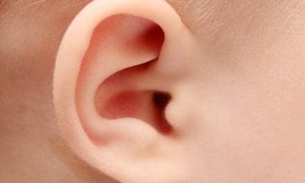
Diehard Beach Boy fans would cringe at the thought of listening to a vintage vinyl album on an old-fashioned hi-fi with a roll of quarters duct-taped to the needle. Although you might recognize the tunes being played, the notes would sound distorted, and the record would likely be scratched beyond repair, notes the National Institute on Deafness and Other Disorders (NIDCD), Washington.
Biophysicists have a similar problem when they study the mechanical properties of tissues in the inner ear. The current technique, called atomic force microscopy (AFM), uses a microscopic probe (like the record player’s needle) on the end of a cantilever (like the arm that holds the needle) to apply force to a material to tell if it’s stiff or soft, sometimes damaging the tissue in the process. In addition, measurements obtained with the standard AFM procedure don’t provide a realistic picture for inner ear tissue since the tissue under the microscope isn’t probed at the frequencies at which it typically functions, sayd NIDCD.
NIDCD researchers may have solved the dilemma. Richard Chadwick, PhD, and Nuria Gavara, PhD, of NIDCD’s Section on Auditory Mechanics, have implemented a new technique that allows them to study the mechanical properties of tissues at frequencies near the upper reaches of human hearing (about 20,000 hertz, or Hz), which has never been possible before, all while preserving the tissue. The study is published in the June 20 advance online issue of the journal Nature Methods.
Instead of poking the sample, the researchers use high-frequency sound vibrations to tell them if a material is soft or stiff. They have modified their commercial AFM device to oscillate a microscopic bead only tens of nanometers above the surface of the tissue, oscillating at high frequency. Also, the oscillations of the bead are extremelly small, less than 10 nanometers, so the researchers don’t hear a thing. Even though the bead and the tissue are not in direct contact, the tissue surface disturbs the bead oscillation that’s reflected as a change in the bead’s mass. If the tissue is rigid and the bead is hovering close to the surface, the increased mass is at its greatest, and the oscillations slow down, lowering the frequency. If the bead is farther away, the increase in mass is lower, and a smaller frequency shift results. Likewise, if the tissue is soft, there is a reduced increase in mass and a smaller frequency shift.
According to Chadwick, the amount of force the new method exerts on the tissue sample in comparison to the standard method would be like comparing the weights of a penny and a gallon of milk; moreover, the force is exerted at the proper frequency.
“It’s a huge difference, so it’s much better for preserving the sample,” he says.
In addition, the researchers are now able to measure the properties of a tissue sample at acoustic frequencies similar to those of human hearing. Although sound frequencies have been used to study biological materials in the past—a technique called frequency-modulated atomic force microscopy—they could only been used to assess the properties of a single molecule versus an entire tissue sample.
Chadwick and Gavara are applying their new technology to study the tectorial membrane, a complex membrane overlying the sensory cells, called hair cells, in the inner ear. In earlier work, they demonstrated that the tectorial membrane is arranged as a gradient, going from very stiff to very soft along the length of the membrane, which is about a quarter of an inch. Their new method now confirms these properties exist at frequencies similar to that of human hearing.
Chadwick notes that this new method should be useful, not just for studying tissues that detect sound, but for measuring tissues used in the production of sound, such as the vocal folds, which is also a research interest of the NIDCD.
[Source: NIDCD]