Similarly to how a picked lock gives away that someone has broken into a building, the stiffening of a structure surrounding cells in the human body can indicate that cancer is invading other tissue.
Monitoring changes to this structure, called the extracellular matrix, would give researchers another way to study the progression of disease. But detecting changes to the extracellular matrix is hard to do without damaging it.
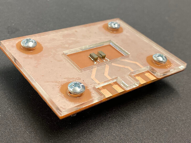
Purdue University engineers have built a device that would allow disease specialists to load an extracellular matrix sample onto a platform and detect its stiffness through sound waves, according to a summary published on the university’s website. The device is described in a study published in the journal Lab on a Chip and demonstrated in a YouTube video at: https://youtu.be/hPvY0Sj0vxY.

A device uses sound waves to detect the stiffness of an extracellular matrix, a structural network that contains cells. Changes in the stiffness of this structure can indicate the spread of disease. (Purdue University photo/Kayla Wiles)
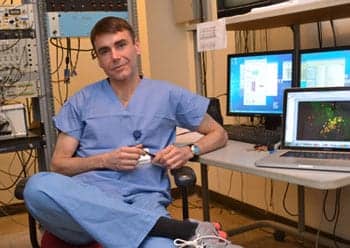
“It’s the same concept as checking for damage in an airplane wing. There’s a sound wave propagating through the material and a receiver on the other side. The way that the wave propagates can indicate if there’s any damage or defect without affecting the material itself,” said Rahim Rahimi, a Purdue assistant professor of materials engineering, whose lab develops innovative materials and biomedical devices to address health care challenges.
Each tissue and organ has its own unique extracellular matrix, sort of like how buildings on a street vary in structure depending on their purpose. The extracellular matrix also comes with “landlines,” or structural and chemical cues, that support communication between individual cells housed in the matrix.
Researchers have tried stretching, compressing or applying chemicals to samples of the extracellular matrix to measure this environment. But these methods also are prone to damaging the extracellular matrix.
Rahimi’s team developed a nondestructive way to study how the extracellular matrix responds to disease, toxic substances, or therapeutic drugs. The initial work for this study was performed in collaboration with the lab of Sophie Lelièvre, a professor of cancer pharmacology at Purdue, to identify how risk factors affect the extracellular matrix and increase the risk of developing breast cancer.
The device is a “lab-on-a-chip” connected to a transmitter and receiver. After pouring the extracellular matrix and the cells it contains onto the platform, the transmitter generates an ultrasonic wave that propagates through the material and then triggers the receiver. The output is an electrical signal indicating the stiffness of the extracellular matrix.
The researchers first demonstrated the device as a proof-of-concept with cancer cells contained in hydrogel, which is a material with a consistency similar to an extracellular matrix. The team now is studying the device’s effectiveness on collagen extracellular matrices.
The device could easily be scaled up to run many samples at once, Rahimi said, such as in an array. This would allow researchers to look at several different aspects of a disease simultaneously.
This research was conducted in Birck Nanotechnology Center in Purdue’s Discovery Park and financially supported by the Breast Cancer Research Program, a Congressionally Directed Medical Research Program (grant W81XWH-17-1-0250).
Original Paper: Zareei A, Jiang H, Chittiboyina S, et al. A lab-on-chip ultrasonic platform for real-time and nondestructive assessment of extracellular matrix stiffness. Lab on a Chip. 2020;20:778-788.
Source: Purdue University, Lab on a Chip
Image: Purdue University/Kayla Wiles


