A Western University team has used the Canadian Light Source at the University of Saskatchewan (USask) in Canada to obtain highly detailed images of the structures in the inner ear responsible for transmitting sound signals to the brain. With these images, they’ve helped pioneer customized programming strategies for cochlear implants.
Because of the cochlea’s tiny, delicate, spiral-shaped structure, and the fact that it is encased in the densest bone in the human body, it is hard to use conventional techniques to study its anatomy and how implants interact with it. Synchrotron imaging changed the game by allowing scientists to visualize the cochlea in incredible detail—roughly at the scale of individual cells.
Further Reading: Researchers Design Tool to ‘Tune’ Cochlear Implants
“We were able to obtain high-resolution data on the synchrotron, and then created beautiful three-dimensional images with our collaborators in Sweden,” says Western University’s Sumit Agrawal, MD.
The team recently published the CLS-enabled mappings of 38 cochleae in the journal Laryngoscope. Agrawal says that this “gold standard data,” based on ultra-detailed imaging of the ear’s anatomy, answers many questions in the field.
Further Reading: Improved Hearing with Optical Cochlear Implants
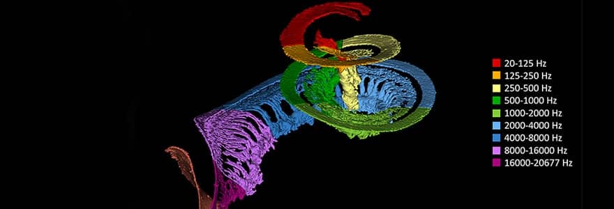
The maps the team created should make a huge difference to the sound quality of cochlear implants. As sound travels down the cochlea, different pitches land at different points in the structure for us to hear them. To tune the sound, an implant needs to match these points for that particular patient’s anatomy. But without a map of the inner ear, cochlear implants can only be “one size fits all.”
“It would be like listening to an out-of-tune piano,” says Agrawal. “What we’re doing now is actually mapping each of the electrodes to tune the piano for each individual patient.”
By combining high-resolution imaging from the Bio-Medical Imaging and Therapy (BMIT) facility at the CLS with the team’s deep learning algorithms, researchers can now create customized maps that match the unique anatomy of each patient’s cochlea. The deep-learning algorithm, too, was partly trained on 3D images produced at the CLS.
Further Reading: Researchers Create Inner Ear Sound Frequency Map
The team has already begun applying this data to patients with cochlear implants, and published a small pilot study using both customized and standard programming. The personally “tuned” implants showed promising results, providing the data needed to proceed with the randomized controlled trial currently underway at Western University and the University of North Carolina.
“The fact that we were able to extract the anatomy from the synchrotron, create a new equation, apply it to patients and improve their performance was really exciting for us,” says Agrawal.
A major grant from hearing implant company MED-EL has helped guarantee the team can keep driving their ambitious synchrotron-enabled research program for many years to come. Announced in late 2023, the $8.5-million donation was matched by Western University, and establishes two endowed research chairs devoted to this kind of independent hearing research.
“Although we have already started applying this translational ‘bench-to-bedside’ research in patients, we are not going to stop using the synchrotron. There are so many other things we can look at, and the synchrotron allows us to see processes in real time,” says Agrawal. “This is just the start.”
Canadian Light Source
The Canadian Light Source (CLS) is a national research facility of the University of Saskatchewan and one of the largest science projects in Canada’s history. More than 1,000 academic, government and industry scientists from around the world use the CLS every year in innovative health, agriculture, environment, and advanced materials research.
The Canada Foundation for Innovation, Natural Sciences and Engineering Research Council, Canadian Institutes of Health Research, the Government of Saskatchewan, and the University of Saskatchewan fund CLS operations.
Featured image: 3D color rendering of synchrotron radiation phase-contrast image of a cochlea, courtesy of Hao Li and Helge Rask-Andersen, Uppsala University Sweden