The VASCilia platform uses deep learning to accelerate imaging analysis by 50-fold, aiding research into hearing loss and gene therapy.
Researchers at the University of California San Diego have developed an artificial intelligence tool that automates the 3D analysis of cochlear hair cells, a process critical for understanding hearing function and developing new treatments for hearing loss. The tool, called Vision Analysis StereoCilia (VASCilia), is described in PLOS Biology and accelerates the imaging process by a factor of 50.
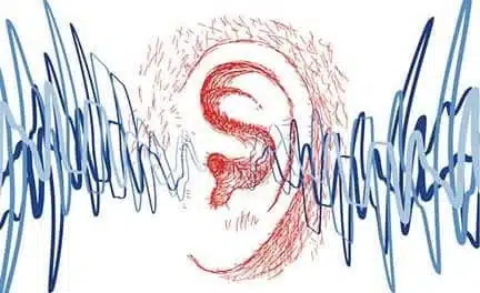
VASCilia provides detailed views of stereocilia, the bundles of protrusions within the cochlea that detect sound. Analyzing the organization and length of these bundles helps scientists understand damage from noise exposure and aging, but the process has traditionally been manual and time-intensive.
“Understanding how stereocilia bundles get disorganized over time, or after exposure to certain environmental stresses, is very important in hearing loss research,” says Uri Manor, a biological sciences assistant professor at UC San Diego, in a release. “We would like to more fully understand how these patterns are disrupted during disease, for hearing research on noise damage and aging.”
The tool is also intended to support the evaluation of gene therapies designed to reverse hearing loss. By providing consistent and accurate quantification of a large number of cells, VASCilia can help measure the effectiveness of such treatments.
“There are children who were born deaf that can now hear because of gene therapy and we expect those treatments for hearing loss to grow,” says Manor in a release. “For gene therapy experiments VASCilia allows us to measure all the cells and we can quantify them very consistently and accurately.”
Developed by a team including postdoctoral scholar Yasmin Kassim, VASCilia uses five deep learning-based models trained on expert-annotated datasets from mice. The platform automates what was previously a slow process of manually interpreting microscopic images of hair cell bundles.
“We’ve reduced the amount of time it takes to analyze the length of these cells by a factor of 50, enabling many additional 2D and 3D quantitative measurements that can be acquired in minutes—work that would otherwise require years of manual analysis,” says Kassim, a computer scientist and a Schmidt AI postdoctoral fellow, in a release. “VASCilia can also detect and quantify subtle patterns of cellular disorganization that are difficult for humans to measure manually.”
The researchers have made VASCilia open-source with the hope of facilitating the creation of a large-scale atlas of cochlea hair cell images to further support advances within the hearing research community.
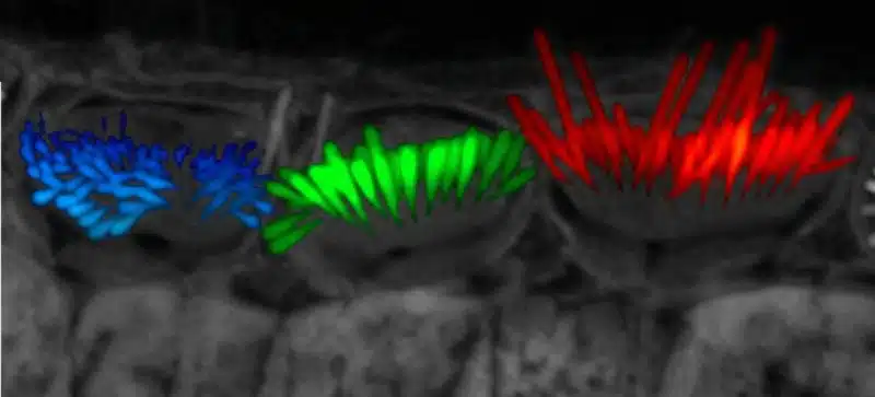
Featured image: Hearing researchers require detailed images of stereocilia, the bundles of protrusions that detect sound and movement. The red hairs featured on the right were rescued to full length due to gene therapy, while green-intermediate hairs received partial treatment and blue received no treatment. Image: Manor Lab, UC San Diego