Bone anchored hearing aids (BAHA™) have been used clinically since 1977, and to date, over 7,000 have been fit. BAHAs are for patients with a conductive hearing loss or a mixed hearing loss where the bone conduction thresholds do not exceed 45 dB (average of 500 Hz, 1000 Hz, and 2000 Hz). That is, a patient may have bone conduction thresholds of 45 dB HL with no measurable air conduction thresholds better than 100 dBHL.
Typical candidates are those with unresolvable middle ear dysfunction (e.g., chronic otitis media) that has not responded to medical or surgical intervention, or congenital facio-cranial conditions such as atresia, Treacher Collins and Goldenhar’s Syndromes. Because a BAHA is still appropriate if there is an additional sensorineural component, these patients would still be candidates even if there was a significant presbycusic or noise-induced component.

|
|
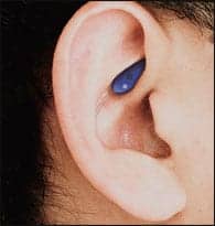
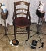
| Fig. 1a-b. Abutment of the BAHA protuding from the skin. | |
The electro-mechanic “receiver” of the BAHA is implanted in the mastoid bone behind the ear (Fig. 1). A small abutment protrudes through the skin. The receiver is not conventional in that it is a small bone conduction device that serves to transduce sound from the external sound processor (the BAHA) to the implanted titanium screw, which is held tightly in the bone and therefore makes an excellent conductor for vibration that travels to the cochlea in a normal bone-conduction fashion. More information can be found in Chasin.1
An advantage of a BAHA over conventional air conduction hearing instruments is that nothing needs to be placed in the ear canal. This minimizes the incidence of repeated ear infection in the case of chronic unresolvable otitis media, while allowing efficient conduction of sound in the case of those with atresias or malformed outer and/or middle ears. For those using conventional bone conduction hearing aids, the use of a BAHA is designed to allow more sound energy, especially in the mid and high frequencies, to be transduced. The use of a BAHA has been shown to improve the articulation index (proportion of audible speech cues) by 18-20% over a well-fit conventional bone conduction hearing aid.2
Clinical Assessment
The question of clinical assessment of this type of device naturally arises. In contrast to an air conduction hearing instrument, the gain and output of a BAHA is independent of the individual’s ear canal characteristics. Standard test procedures involving real ear measurement (REM) are obviously not possible; nor is the use of a 2cc coupler or other volume-related ear simulator. This article details the correct use of some sound field measures including functional gain measurements and the pitfalls to be avoided during the evaluation of benefit for the BAHA. This technique can also be used to assess conventional bone conduction hearing aid fittings.
The most obvious alternative to REM testing is functional gain testing. Functional gain, as the name suggests, is a measure of the improvement (gain) that an individual receives from the device, with measurment based on responses from the individual. Typically, a patient is placed in a sound field and asked to respond to a stimulus with and without the hearing aid(s) in place. The resulting difference is “calibration independent” in that all calibration issues are identical for both aided and unaided conditions, with the result that they are subtracted off. The result, like insertion gain, is expressed in dB (not dB SPL or dB HL). As long as the patient has not changed position, and other signal generation issues have not been encountered, the difference in decibels can be added directly to the audiogram. This would be the same as REM testing—the insertion gain can be added directly to the audiogram, and indeed, most REM manufacturers use software that allows the results to be expressed either in SPL or as an aided audiogram in HL calibration.
Well-known potential clinical problems of functional gain measurement are related to:
1) Not being able to generate a sufficiently intense stimulus for the unaided portion of some measurements;
2) Using inappropriate stimuli that are neither narrow-band enough to be frequency specific, nor wide-band enough to prevent standing waves; and
3) The possibility that internal masking of a person’s own auditory system does not affect audibility of low-frequency, low-intensity stimuli.
However, even if the above technical problems are resolved, erroneous results for some patients can be obtained. The first issue concerns the non-test ear response. For example, in a case of unilateral conductive hearing loss, or when the non-test ear is significantly better than the test ear, the stimulus may be heard by the non-test ear. Clinicians have used masking through earphones placed over the non-test ear, and earplugs that fit in the non-test ear. Sufficient masking, as long as overmasking does not occur, is valid. However, the use of ear plugs in the non-test ear may not be sufficient. Even a deeply seated ear plug will only attenuate the lower frequencies by 25 dB (and the higher frequencies by 35-38 dB), and, depending on the hearing loss, this may not be sufficient. Understandably, given that we are dealing with conductive and mixed hearing losses, this would be more of an issue in the lower frequency region.
A second related issue involves the 2000 Hz region where the attenuation of the human skull is only about 40 dB (see Berger3). Sounds that are more intense than 37-40 dB SPL can enter the skull and, via bone conduction, bypass the ear canal and go directly to the cochlea. This is why hearing protection cannot yield more than 40 dB attenuation in the 2000 Hz region regardless of the attenuation measured with any of the various “microphone in the ear canal” testing methods.
Clinically, the hearing care professional needs a frequency-specific assessment procedure that can be used on all hearing losses, including those losses that warrant the use of a BAHA. The solution involves recognizing that whereas the unaided portion of the functional gain measurement may be affected by many artifacts, the aided portion is relatively free of artifacts. Sound field standards like the ANSI-1996, S3.64 have (in part) resolved this problem, but not all audiometric facilities use the newer standard. Many facilities do not routinely calibrate their sound fields either because of financial constraints, or because the use of REM obviates the need for a calibrated sound field. A clinically expedient (10 minute) calibration technique is proposed that can be used to write frequency-specific aided results on the audiogram (in HL calibration). The essence is similar to the 1996 ANSI sound field standard.4
Calibrating the Sound Field: The procedure for calibrating the sound field in order to perform bone conduction assessment is relatively straightforward:
1) Set the sound field stimulus to warble or narrow band noise at a 70 dB dial reading.
2) Measure the SPL at the location of the center of the head as if the patient were seated in the room (without the patient).
3) Record the SPL and subtract the MAF (Minimal Audible Field) values shown in Table 1 from the frequency-specific SPL dial readings.
4) Subtract the 70 dB dial reading from the resulting number in order to get a value that you can write on your audiogram (i.e., equivalent HL).
For example, if you measure an SPL at 1000 Hz of 65 dB SPL (with a dial reading of 70 dB), there is a -5 dB difference. You need to subtract another 4 dB (see Table 1) since that is the MAF correction. The real correction is therefore –9 dB (= -5dB plus -4 dB). You only need to do this once. Once calibrated, in this example, an aided result on your dial of 30 dB is actually 21 dB HL. This can be written directly on the audiogram.
|
Frequency (Hz) |
250 | 500 | 1K | 2k | 3k | 4k |
| Measured SPL Dial reading MAF correction (0°) (Equiv. to dB HL) |
13 | 6 | 4 | .5 | -4 | -4.5 |
| Table 1. Worksheet for the conversion of audiometer dial readings to equivalent dB HL. The “Dial reading/Measured SPL” only needs to be done once for initial calibration. The calibration takes approximately 10 minutes with Measured SPL – Dial Reading – MAF Correction = Equivalent dB HL (refer to text for full explanation). | ||||||
|
Frequency (Hz) |
250 | 500 | 1K | 2k | 3k | 4k |
| MAF correction (0°) | 13 | 6 | 4 | .5 | -4 | -4.5 |
| MAF correction (45°) | 12 | 3 | 0 | -2.5 | -9 | -8.5 |
| Table 2. Frequency-specific calibrations from ANSI (1996) for 0° azimuth and 45° azimuth. Note large differences in the higher frequencies. | ||||||
In the case of BAHAs or conventional hearing instruments, this aided result can be compared with the bone conduction threshold at that frequency. Sound field calibration is a function of azimuth angle of signal delivery. Table 2 shows the MAF corrections for both 0° azimuth (also shown in Table 1) and the corrections for a 45° angle. (With conventional bone conduction hearing aids, complete closure of the “air/bone gap” may not be the target, since audiometric bone conduction oscillators, as well as those used for conventional bone conduction hearing aids, may have different characteristics).5
Sound Field SPL: There are two quick methods to determine the exact sound field SPL reading. One is with the use of a sound level meter (C-weighting) and the other is with a REM system. When using a REM system for this purpose, one must first disable both the reference microphone and the speaker. Various manufacturers have differing methods to accomplish this, but once performed, the REM system essentially becomes an in-situ sound level meter, and the peak value on the screen display will be an accurate measure of the peak spectrum of the stimulus. For example, Audioscan requires that the stimulus level be set to 0 dB, and the Frye system requires that the stimulus be turned “off.” In both of these cases, the REM system will measure external stimuli such as a warble, and the reference microphone will not alter the measurement.
The Skull Simulator & Functional Gain
Peder Carlsson6 developed a skull simulator, which in essence is a modified artificial mastoid. The BAHA uses a small “point source” screw that is embedded in the mastoid bone. This point source approach is designed to be an efficient mechanical transducer that yields significantly more gain (+20 dB) than conventional bone conduction hearing aids. This is primarily why the BAHA has been reported to be useful for a wider range of hearing losses than conventional bone conduction hearing aids. Such a skull simulator can be used to measure the electro-mechanical characteristics of the BAHA for both quality control concerns and to develop “coupler”/functional gain transfer functions.
These functions will be limited by the accuracy of the functional gain measure—a standard error for 7 dB or 5 dB increment steps and 2.8 dB for 2 dB increment steps. Because a BAHA has a well-defined connection that has stable electro-mechanical characteristics, such a transfer function is stable and can be applied directly to specification sheets to calculate expected functional gain in an a priori manner. It should be pointed out that this would not be the case with the now discontinued Xomed Audiant that used a less efficient “bone conductor-like” transducer.
Usefulness of Assessment
Functional gain, in conjunction with the appropriate skull simulator, can yield data that is both reliable and valid. Frequency-specific information can be determined within reasonable clinical time requirements to verify or to make adjustments. The data obtained can be correlated with other subjective test results that many clinicians have been using over the years, including the signal-to-noise ratio at 50% correct7, various speech-in-noise tests, and client satisfaction scales such as the SADL.8 Functional gain testing has some limitations, but these can be minimized if the incremental step size is kept small (e.g., 2 dB), especially near threshold, and as long as care is taken to prevent errors from entering into the calculations.
Acknowledgements
The author wishes to thank Elliott Berger for his comments on an earlier draft of this paper.
This article was submitted to HR by Marshall Chasin, MSc, coordinator of research at the Canadian Hearing Society. Correspondence can be addressed to HR or Marshall Chasin, Canadian Hearing Society, 271 Spadina Road, Toronto, Ontario M5R-2V3, Canada; email: [email protected].
References
1. Chasin M: Current trends in implantable hearing aids. Trends in Amplification 1997; 2 (3).
2. Chasin M: Bone conduction implants: The when and why. Hearing Review. 2000; 7(12):56-58.
3. Berger EH: Methods of measuring the attenuation of hearing protection devices. J Acoust Soc Amer 1986; 79 (6): 1655-1687.
4. American National Standards Instute. ANSI S3.6 (1996): Standard for audiometers. New York: ANSI, 1996.
5. Stenfelt S: Hearing by bone conduction: Physical and physiological aspects. Goteborg, Sweden: Dept. of Signals and Systems, Medical Electronics Group: Chalmers Univ of Technology, 1999.
6. Carlsson PU: On Direct Bone Conduction Hearing Devices. Technical Report No. 195, Goteborg, Sweden: Chalmers Univ. of Technology, 1990.
7. Hakansson B, Liden G, Tjellstrom A, Ringdahl A, Jacobsson M, Carlsson P & Erlandson BE: Ten years of experience with the Swedish bone-anchored hearing system. Ann Otol Rhinol & Laryngol 1990; Suppl. 151, 99 (10): Part 2.
8. Cox RM & Alexander GC: Measuring satisfaction with amplification in daily life: the SADL scale. Ear & Hear 1999; 20: 306-319.