Patient Care | February 2023 Hearing Review
A deep dive into the next five year, P50 clinical research grant on cochlear synaptopathy.
By Kathryn Sutherland

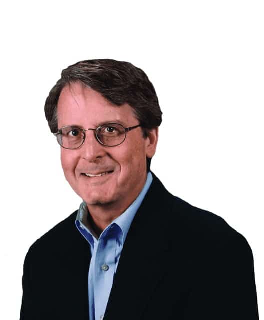
I recently had the pleasure of speaking with Charles Liberman, PhD. Who, in August of 2022, lead a team of researchers at the Mass Eye and Ear, where the team was awarded a second, five-year, $12.5 million P50 Clinical Research grant to continue their research on cochlear synaptopathy. We discussed his background, including how he became interested in the study of hearing, research findings on cochlear synaptopathy, and a preview of research to come in the next five years.
Dr Liberman earned his PhD from Harvard University in 1976 before joining Mass Eye and Ear as a research associate and was appointed director of the Eaton Peabody Laboratories in 1996, which has undergone immense growth under his leadership.
In 2009, Drs Liberman and Sharon Kujawa, PhD, uncovered a new type of inner-ear damage called cochlear synaptopathy, also known as “hidden hearing loss.” Their landmark discovery, which has changed how the scientific community understands hearing loss, showed that there could be permanent damage to auditory nerve fibers from loud noise or age long before there is damage to the sensory cells.
Sutherland: Welcome, Dr Liberman, and congratulations on being named the [2022 La Fondation Pour l’Audition grand prize recipient]. Would you please share your background with our readers?
Liberman: I became interested in how the brain works as a senior at Harvard College after taking a neurobiology course.
Music was also very important to me, and these two interests came together for me in the study of hearing. There weren’t any courses at Harvard on hearing, so I sought advice on whom I should see and was told to connect with Nelson Kiang, the Director of the Eaton-Peabody Laboratory of Auditory Physiology, at the Massachusetts Eye and Ear Infirmary. I ended up taking a year- long reading course with him, studying the original literature on hearing and deafness. He encouraged me to become his graduate student. I joined the Eaton-Peabody Labs, and I’ve been at the Mass Eye and Ear ever since.
For my thesis, Dr Kiang suggested that I look at noise damage. This was a long time ago before we knew much about how the ear worked. He suggested that if we used loud sounds to damage the ear, we could create some interesting patterns of damage. For example, perhaps we could selectively destroy one type of sensory cell and not the other and reveal how the system normally works by seeing what kind of dysfunction there is when you destroy certain elements.
However, as my research progressed, I got more interested in noise damage per se. Why is it that overexposure to sound damages hearing? Why is it that threshold elevation is sometimes reversible and other times irreversible? What’s really going on inside the ear? Where is the damage? What are the sensitive elements? And what are the weakest links?
Since that time, I’ve continued, in one form or another, studying noise damage and other types of cochlear insults that cause sensorineural hearing loss, like ototoxic drugs and aging. I’ve also continued to study aspects of the normal auditory system. How are sounds converted into electrical signals in the auditory nerve? How is that information packaged and sent to the brain in the normal ear? How does that coding of acoustic information become degraded or altered in sensorineural hearing loss? And how do those insights help us understand the range of hearing impairments associated with different types of sensorineural loss, such as problems hearing noise or tinnitus?
Sutherland: Fascinating subjects, with many different yet related questions to answer. It sounds like a puzzle.
Liberman: The first decades of my research focused on animal models of sensorineural hearing loss. In the past ten years, my research has turned more to human subjects. In collaboration with Dr Stéphane Maison here at Mass Eye and Ear, we are studying living humans who have varying degrees of hearing loss. In addition, my own research group has spent a lot of time looking at tissue damage in the inner ears from human autopsy specimens.
Sensorineural hearing loss happens in the inner ear, where the sensory cells turn mechanical vibrations into electrical signals and the auditory nerve fibers take that information to the brain. The reason it’s called sensorineural hearing loss is that it damages either the sensory cells or the neurons of the inner ear. In a living human, you cannot see the structures of the inner ear with cellular-level resolution. So, if you want to see what’s damaged in the inner ear of humans at present, the only option is to look at autopsy specimens.
I am fortunate that here, at Mass Eye and Ear, scientists have been collecting autopsy specimens for close to 70 years, soliciting donors to donate their tissue after death, and then harvesting the part of the skull that has the inner ear in it, processing it and preparing it for study and archiving the tissue. Then, over the last ten years or so, we’ve reinvestigated a lot of that archive to see if the new insights we gained in the animal models of noise-induced hearing loss and age-related hearing loss translated to the human.
Sutherland: Can you explain how you were able to use the archival tissue to conduct your studies?
Liberman: The Mass Eye and Ear collection includes more than 2000 inner ear specimens that have been sectioned and mounted on microscope slides for archival analysis. Many of these cases also have audiometric data acquired within a few years of death, along with detailed medical and otologic histories. Although most of these cases have been studied in the past, our approach has been to quantitatively assess the nature and degree of tissue damage across large numbers of cases and apply statistical modeling to tease out the functionally important structural changes underlying sensorineural hearing loss.
Sutherland: How has the study of human tissue evolved?
Liberman: We started processing newly acquired material in different ways that gave us additional insights into the extent of nerve damage in the aging ear. A longstanding dogma in the study of sensorineural hearing loss was that the sensory cells are the most vulnerable elements and that the nerve fibers degenerate only after the sensory cells are destroyed. We showed that the reverse was true. As we age, our auditory nerve fibers disconnect from the sensory cells long before the sensory cells disappear.
This is important because nerve fiber loss does not affect the audiogram, the classic test of hearing function, until it reaches 80 or 90%. The audiometric thresholds, the sound level required to detect a tone, are fundamentally determined by the health of the sensory cells. However, we believe that neural loss limits intelligibility of sounds and that this loss of auditory nerve fibers, which “hides behind the audiogram,” is a major cause of the problems hearing in noise that are so characteristic of age-related hearing loss, as well as many other types of sensorineural hearing loss.
Sutherland: What you’re working on will revolutionize how audiologists test for hearing. I know that you have now been awarded a new P50 grant to continue the next five years of this study. Is there more of a pinpoint focus that you will be working on specifically to replace an audiogram? Or are you working on other ideas for testing outside of just the gold standard for hearing testing?
Liberman: Yes, one of the main focuses of our collaborative grant is to develop and validate different tests of neural degeneration in the inner ear. The human work includes two arms. One carries out electrophysiological and behavioral tests on subjects designed to assess the degree of neural degeneration and compares those results with the performance on difficult word-recognition tasks. The other component looks at human autopsy specimens to assess sensory cell and neural degeneration in different types of sensorineural hearing loss. Then, it compares those data to the word scores measured premortem. This human work is complemented by animal studies, where we study animals with sensorineural hearing loss, applying many of the same tests we apply in humans and then removing the inner ears to directly measure loss of sensory cells and nerve fibers.
Sutherland: It sounds very in-depth, with a community of researchers involved.
Liberman: Good strategies for making progress.
Sutherland: Let’s talk about tinnitus. As we know, there are 50 million people suffering from varying degrees of tinnitus. It sounds like your work will provide insights into the causes and possibly bring about some type of treatment or therapy that might help people suffering from tinnitus.
Liberman: Yes, absolutely. Comparing people with and without tinnitus is a big part of the next five years of our combined work together. Although there are likely many ways to cause tinnitus, we believe that, in a lot of cases, tinnitus is elicited by nerve degeneration in the inner ear. There is accumulating evidence from animal studies that if you have less neural activity going to the auditory brain, the central circuits try to compensate by jacking up the gain. This increased gain leads to a number of problems, the most important being tinnitus and hyperacusis, reduced tolerance for moderate-level sounds.
It’s important to understand the underlying mechanisms, but, of course, what really matters is the development of treatments. In another collaborative grant, I work with Dr Gabriel Corfas of the University of Michigan on the development of approaches to neural regeneration. What’s exciting and promising about the problem of “hidden hearing loss” is that the neural degeneration in the aging ear disconnects the auditory nerve from the hair cell but not from the brain. The survivability of auditory nerve fibers, even in the face of massive degeneration of sensory cells, is why cochlear implants work.
In “hidden hearing loss”, we have a situation where a lot of the sensory cells survive and a lot of the neurons survive, but they’re disconnected. We have shown in mice that, after noise damage, you can stimulate the auditory nerve fibers to send out new processes and make new connections with the hair cells by delivering a naturally occurring molecule called neurotrophin-3. We have also shown in the aging mouse that we can halt the progressive loss of these neural connections by using genetic tools to cause the overproduction of neurotrophin-3 in the inner ear.
Sutherland: I’m sure that our readers are going to be very interested to learn the results of this next phase of research. How close do you feel you are to be able to revive the connection?
Liberman: Yes, the “When” question is a hard one. We believe these treatments are applicable in principle to humans, but there is a lot of further work to be done before such treatments might appear in the clinic. In the animal model, the neurotrophin delivery treatment only works if applied within a few days of the noise exposure and that presents obvious practical challenges in the human context. It’s possible that using gene therapy approaches, we can chronically boost the levels of neurotrophins in the inner ear to improve the survival of auditory nerve connections in aging and after noise. We have proof of principle experiments in an animal model, but there’s still a while to go before it’s going to be applicable in the clinics.
Sutherland: We have a few minutes left of our time together. Do you have anything else you would like to add to our conversation?
Liberman: We’ve pretty much covered the bases.
Sutherland: Thank you, Dr Liberman. Take care.
Liberman: You too.
This interview has responses that have been condensed and/or edited for readability.


