Novel, fully digital, high-resolution positron emission tomography/computed tomography (PET/CT) imaging of small brain stem nuclei can provide clinicians with valuable information concerning the auditory pathway in patients with hearing impairment, according to a new study published in the March issue of The Journal of Nuclear Medicine. A summary of the study is available on the Society of Nuclear Medicine and Molecular Imaging (SNMMI) website. Using 18F-FDG PET/CT imaging, researchers found that patients with asymmetrical hearing loss have reduced glucose metabolism in parts of the brain stem and primary auditory cortex. The latter may be influenced by cortical reorganization and thus, hopefully help to predict the chance that a cochlear implant will improve hearing.
“With the possible exception of few dedicated high-resolution research scanners, earlier PET/CT systems with lower resolution did not permit clear-cut identification and assessment of brain stem nuclei,” said Iva Speck, MD, resident of otorhinolaryngology at the University of Freiburg Medical Center in Freiberg, Germany. “Today, the use of fully digital clinical PET/CT systems permits greatly enhanced imaging and quantitative assessment of small brain stem nuclei, such as the inferior colliculus (IC), the part of the midbrain that acts as a main auditory pathway for the body.”

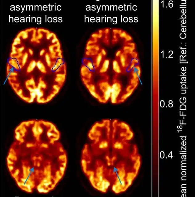
In the study, 13 patients with asymmetric hearing loss underwent 18F-FDG PET/CT imaging. The scans were reviewed by two experienced readers who examined regional glucose metabolism in the IC and the primary auditory cortex (PAC)—a part of the brain known to undergo metabolic changes based on acoustical outside input and transformation to neuronal signals from the cochlea hair cells to the auditory nerve fibers. The readers rated the scans as to whether glucose metabolism showed no asymmetry or mild, moderate, or strong asymmetry to the left or to the right for the IC and PAC separately. Statistical analyses were performed to determine the effect of the duration of hearing impairment on glucose metabolism and to compare glucose metabolism between the IC and PAC.
Regional glucose metabolism of both the IC and PAC was significantly reduced on the contralateral (opposite) side of the poorer-hearing ear, as compared to the ipsilateral (same) side. In addition, a longer duration of hearing impairment was associated with a higher metabolism on the contralateral PAC. By contrast, duration of hearing impairment did not predict regional glucose metabolism for the ipsilateral PAC or either side of the IC.
“Previous studies suggest that the association between longer duration of hearing impairment and higher glucose metabolism indicates cortical reorganization. In bilateral deaf patients this has been shown to lessen the benefits of cochlear implants,” said Speck. “Prediction of a successful cochlear implant outcome might benefit from improved imaging with fully digital PET/CT systems, as large parts of the auditory system, including small brain nuclei such as the IC, can be assessed for preoperative patient characterization.”
She continued, “Beyond this topic, the study’s findings are of interest for other neurological research fields, like neurodegenerative diseases, which often affect brain stem nuclei early in disease course. Digital PET pushes the limits of what can be imaged and contributed to patient care by molecular imaging.”
Original Paper: Speck I, Arndt S, Thurow J, et al. 18F-FDG PET Imaging of the Inferior Colliculus in Asymmetric Hearing Loss. The Journal of Nuclear Medicine. 2020;61:418-422.
Source: Society of Nuclear Medicine and Molecular Imaging, The Journal of Nuclear Medicine
Image/Media: Iva Speck, University Hospital Freiburg, Germany, SNMMI, YouTube